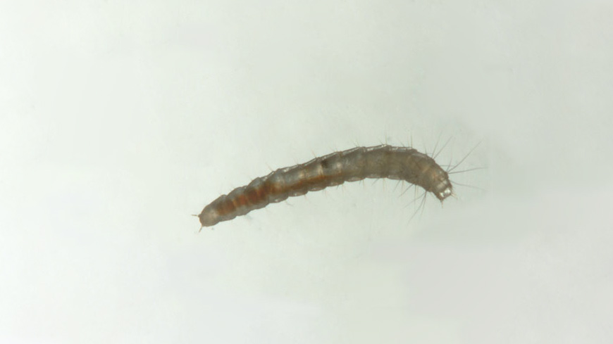
Img 1 Picture of a cat flea larva (Ctenocephalides felis) on a light-colored background.
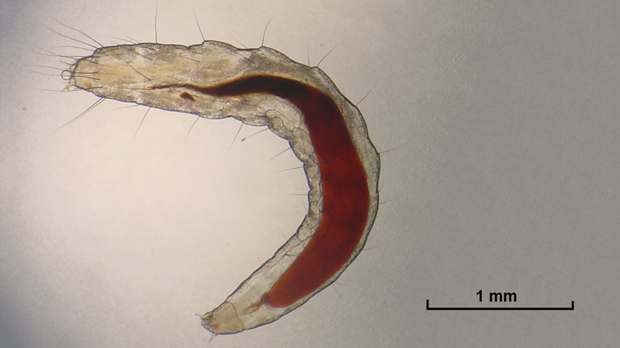
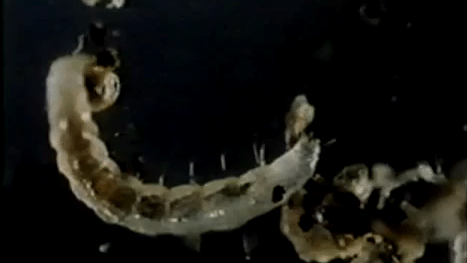
Img 2 Microscope image of a cat flea larva (baby flea) with visible red blood in its digestive track.

Img 3 Illustration of a cat flea larva compared to the size of a sharp #2 pencil tip (1 mm).

Img 4 Picture of shed larval casings from previous moults, along with an egg and feces from adults.
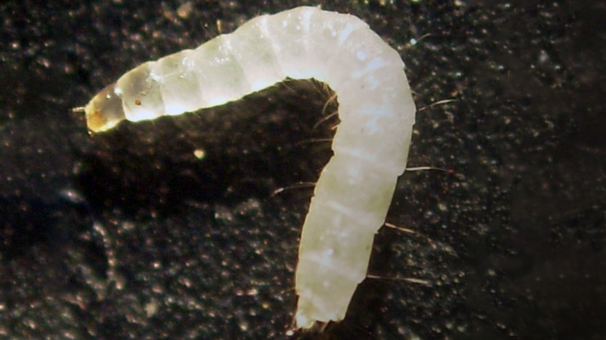
Img 5 Picture of 3rd instar cat flea larva in the prepupa stage after voiding its gut.
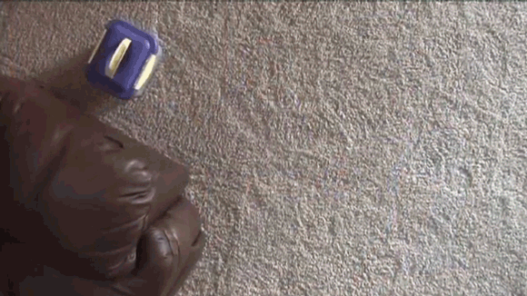
Img 6 Flea larvae living within carpet fibers, the most common place to find larvae in homes.
Img 7 Cat flea larva hatching from its egg with an egg burster spine located on its head.
Img 8 Cat flea larvae feeding on a spherule of adult fecal blood (“flea dirt”).
Img 9 A cat flea larva creating a silken cocoon. The mandibles are used to form the structure while silk is excreted from labial (salivary) glands.


You must log in to post a comment. Log in now.